Bio-optical Imaging Systems
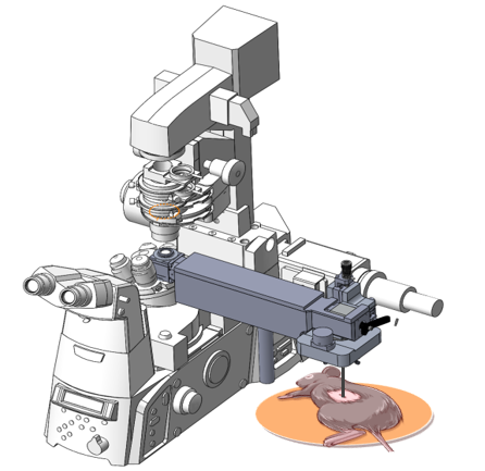
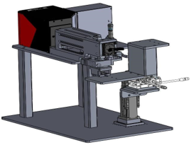
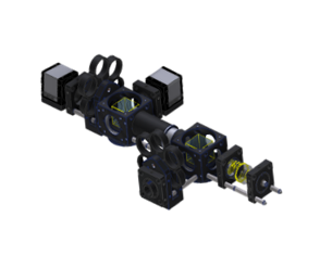
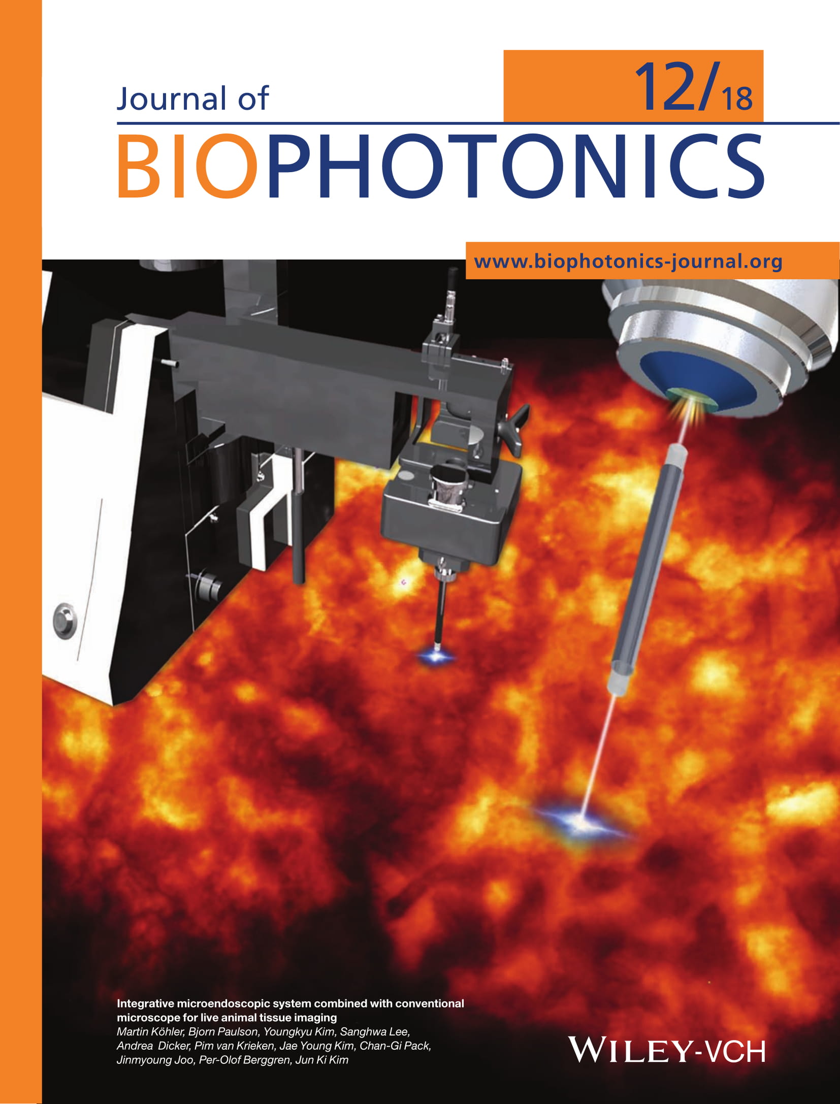
Microscope-attachable endoscopy systems
While microscopes are ideal for seeing biological tissues, microendoscope probes are ideal for accessing organs and tissues in vivo. Our custom-engineered optical relays seamlessly connect endomicroscope probes to pre-existing widefield and confocal microscopes, allowing the imaging power of the microscope to be brought to in vivo biological questions. Our specialized designs allow endomicroscope probes to rotate, switch between vertical and horizontal orientations and adapt between the microscopes of a wide variety of manufacturers, allowing the most effective use of both front- and side-view endoscope probes.
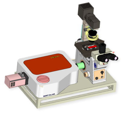
Intravital Confocal/Multiphoton laser scanning systems
Imaging narrow sections of a thicker biological tissue is necessary for the investigation into cellular processes. Confocal laser scanning systems make this possible by maximizing brightness and minimizing otherwise prohibitive noise. We incorporate these systems into endoscopes to obtain high-quality micrographs from the internal organs of living animals.
Super-Resolution Radial Fluctuations (SRRF) system
Achieving resolution beyond the Abbe limit of light diffraction has recently become possible without need for complex illumination, specially selected fluorophores, and toxic light intensities. Our laboratory is equipped with a state-of-the-art SRRF super-resolution system for live-cell imaging.
Combined bright field – fluorescence imaging system
Improved optical diagnosis is possible when clinicians are provided with a combination of data from bright-field and fluorescence imaging modalities. We design proximal optical systems which make multiple imaging modalities possible through existing endoscopic probes.
Raman spectroscopy system
Advanced diagnosis is possible when molecular markers of disease are characterized by Raman spectroscopy in vitro and in vivo. Recently Raman spectroscopy for all-optical diagnosis has demonstrated speed and accuracy compatible with clinical use. With our Raman spectroscopy system, we are pursuing the use of advanced techniques to make diagnoses possible with significantly reduced sample sizes and trauma.
Microscope-attachable endoscopy systems
While microscopes are ideal for seeing biological tissues, microendoscope probes are ideal for accessing organs and tissues in vivo. Our custom-engineered optical relays seamlessly connect endomicroscope probes to pre-existing widefield and confocal microscopes, allowing the imaging power of the microscope to be brought to in vivo biological questions. Our specialized designs allow endomicroscope probes to rotate, switch between vertical and horizontal orientations and adapt between the microscopes of a wide variety of manufacturers, allowing the most effective use of both front- and side-view endoscope probes.
Intravital Confocal/Multiphoton laser scanning systems
Imaging narrow sections of a thicker biological tissue is necessary for the investigation into cellular processes. Confocal laser scanning systems make this possible by maximizing brightness and minimizing otherwise prohibitive noise. We incorporate these systems into endoscopes to obtain high-quality micrographs from the internal organs of living animals.
Super-Resolution Radial Fluctuations (SRRF) system
Achieving resolution beyond the Abbe limit of light diffraction has recently become possible without need for complex illumination, specially selected fluorophores, and toxic light intensities. Our laboratory is equipped with a state-of-the-art SRRF super-resolution system for live-cell imaging.
Combined bright field – fluorescence imaging system
Improved optical diagnosis is possible when clinicians are provided with a combination of data from bright-field and fluorescence imaging modalities. We design proximal optical systems which make multiple imaging modalities possible through existing endoscopic probes.
Raman spectroscopy system
Advanced diagnosis is possible when molecular markers of disease are characterized by Raman spectroscopy in vitro and in vivo. Recently Raman spectroscopy for all-optical diagnosis has demonstrated speed and accuracy compatible with clinical use. With our Raman spectroscopy system, we are pursuing the use of advanced techniques to make diagnoses possible with significantly reduced sample sizes and trauma.
* Selected papers :
1.“Mitotic Chromosomes in Live Cells Characterized Using High‐Speed and Label‐Free Optical Diffraction Tomography” Cells , 8(11), 1368, Oct, 2019
J. Biophotonics
* Selected papers :
J. Biophotonics