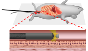
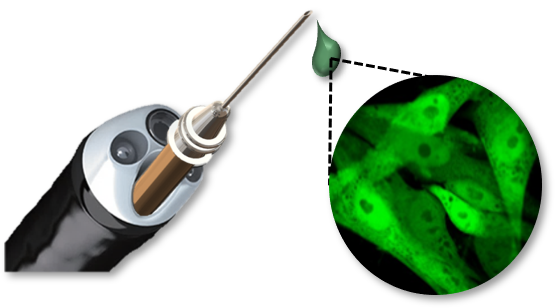
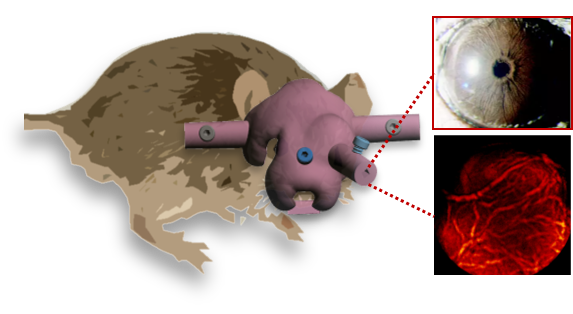
Specialized software and manufacturing processes allow our lab to design custom optical systems, including imaging lenses, lens assemblies, and optical probes. For the purpose of imaging biological systems in vivo, we design rigid endoscopic probes based off the use of gradient index (GRIN) lenses with diameters down to 0.350 mm. Where access to tissues is restricted by organ curvature, we also design flexible endoscopic probes based on coherent fiber bundles. Together, these allow the clear imaging of a wide variety of organs and tissues.